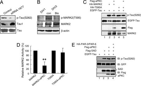
Fig. 2.
MARK2-mediated tau phosphorylation at S262 is inhibited by aPKC. (A) Primary neurons were transfected with control or siRNA-1677 and cultured for 72 h. Cell lysates were subjected to immunoblotting with antibodies against p-tau (S262), Tau1, or total tau. (B) Hippocampal neurons at DIV1 or DIV3 were treated without or with 5 μM bisindolylmaleimide (Bis) for 7 h. Cell lysates were subjected to immunoblotting with indicated antibodies. (C) Lysates of HEK-293 cells transfected with EGFP-tau alone or together with indicated plasmids were subjected to immunoblotting with antibodies against p-tau (S262) or individual tags. (D) Quantitative analysis of data in C. The relative amount of tau (S262) against total tau was normalized to represent MARK2 activity, with that from MARK2 or T595A transfection alone as 100%. Data are shown as means ± SEM (n = 3; ∗∗, P < 0.01). (E) Lysates of HEK-293 cells transfected with indicated plasmids were subjected to immunoblotting.

