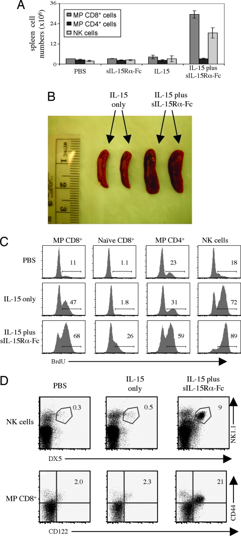
Fig. 3.
Soluble IL-15Rα augments IL-15-mediated host lymphocyte proliferation. (A) Normal B6 mice were injected i.v. on days 1 and 2 with PBS, sIL-15Rα-Fc alone, IL-15 alone, or sIL-15Rα-Fc/IL-15 as described for Fig. 2A. Total numbers of CD8+ MP T cells, CD4+ MP T cells, and NK cells recovered from spleen on day 3 are shown. (B) Spleens from A were photographed as indicated. (C) Mice were treated as in A, except that the mice also were given an i.v. injection of BrdU on day 1 and placed on BrdU in the drinking water until killing. Shown is BrdU staining for MP CD8+, naïve CD8+, MP CD4+, and NK cells. (D) IL-15Rα–/– mice were injected i.v. on days 1, 3, 5, and 7, with PBS, IL-15 (0.6 μg), or IL-15 (0.6 μg)/sIL-15Rα-Fc (3 μg). The data show staining of spleen cells on day 9. For B–D, representative data are shown. All data are representative of at least two independent experiments.

