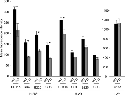
Fig. 2.
Splenocytes from ERAP1−/− mice express lower cell-surface MHC class I than those from ERAP1+/+ mice. Splenocytes from ERAP1+/+ mice (WT, black bars) or ERAP1−/− mice (KO, gray bars) were stained with mAb to H-2Kb or H-2Db or, as a control, to I-Ab and various antibodies to distinguish splenocyte subsets (CD11c, predominantly dendritic cells; CD8+and CD4, predominantly T lymphocyte subsets; B220, predominantly B cells) as indicated on the x axis. Shown are mean fluorescence intensities with background staining by an irrelevant antibody subtracted (averages of three mice; error bars represent 1 SD; representative of five experiments). Asterisks indicate statistically significant differences (P < 0.05, Student t test) between ERAP1+/+ and ERAP1−/− splenocytes.

