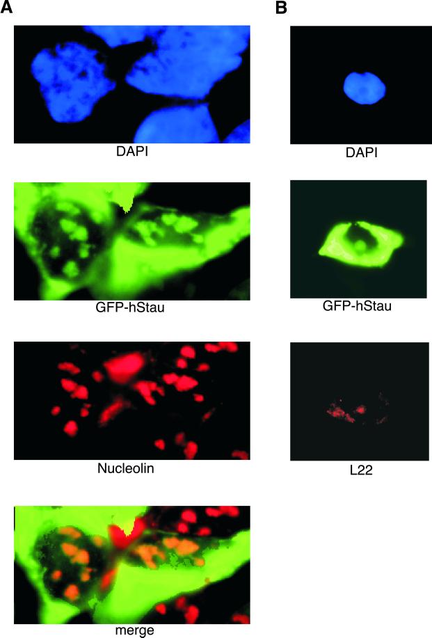
Figure 5.
Subcellular localization of the hStau and the L22 protein. (A) Green fluorescence of GFP-hStau and nucleolin immunofluorescence of 293 cells expressing the GFP-hStau fusion. Cells were stained with DAPI (blue, top), green fluorescence (green, second from top), and anti-nucleolin (red, second from bottom), and images were merged (bottom). (B) Green fluorescence of GFP-hStau and L22 immunofluorescence of 293 cells expressing the GFP-hStau fusion. Cells were stained with DAPI (blue, top) and L22 antiserum (red, bottom) and processed for a green fluorescence image (green, middle).

