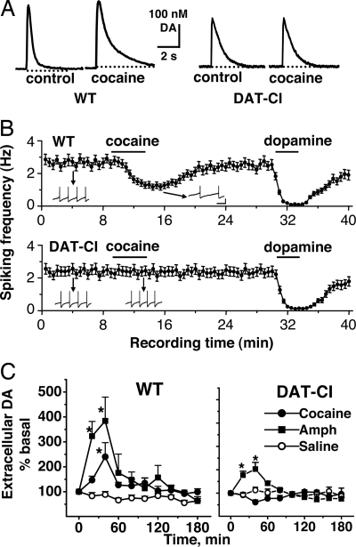
Fig. 3.
Differential effects of cocaine on DA release, uptake, and DA neuron firing between genotypes measured in vitro and in vivo. (A) FCV was used to measure DA release and reuptake in accumbal slices in the absence or presence of 4 μM cocaine. Cocaine prolonged the DA decay time from brain slices of WT mice but not of DAT-CI mice. (B) The frequencies of spontaneous action potentials in DA neurons were recorded with whole-cell patch clamp and presented as mean ± SEM. The effects of bath application of 5 μM DA and 5 μM cocaine are shown. Cocaine significantly decreased the frequency of DA neuron firing in WT mice but not in DAT-CI mice. Insets show actual spike traces. Spikes were truncated for display. (Scale bars: 500 ms and 20 mV.) (C) Extracellular DA in the NAc was assessed by microdialysis in free-moving mice. The average of the two samples before the treatment (i.p.) of cocaine (20 mg/kg), AMPH (2.5 mg/kg), or saline was used as the baseline (100%, 0 min). Cocaine significantly increased extracellular DA level in the NAc of WT mice (F1,113 = 28.4; P < 0.0001) but not in DAT-CI mice (F1,113 = 2.89; P > 0.05). AMPH elevated extracellular DA level in both WT (F1,81 = 52.0; P < 0.0001) and DAT-CI (F1,81 = 6.5; P < 0.05) mice. Two-way ANOVAs were performed. Data represent mean ± SEM (n = 5–8).

