Fig. 1.
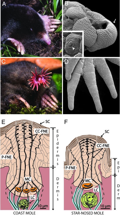
The coast mole (Scapanus orarius) and the star-nosed mole (Condylura cristata) showing adaptations to a fossorial lifestyle. (A) The coast mole has large claws and tiny eyes and lacks an external pinna. (B) A scanning electron micrograph of the coast mole’s snout covered with Eimer’s organs. An arrow marks organs worn by soil abrasion (see Discussion). (Inset) An Eimer’s organ showing the circular disk at the top of the central cell column (arrowhead). (C) The star-nosed mole showing the rhinarium composed of 22 appendages. (D) A scanning electron micrograph of appendages covered with Eimer’s organs from the right lower quadrant of the star. (E) Schematic representation of Eimer’s organ in the coast mole. The epidermis contains a central epithelial cell column associated with intraepidermal free nerve endings (CC-FNE) that course to the stratum corneum (SC). A ring of smaller peripheral free nerve endings surrounds the central column free nerve endings (P-FNE). Merkel cell–neurite complexes (MC) and lamellated corpuscles (LC) reside at the base of each organ. (F) Schematic representation of the smaller Eimer’s organ in the star-nosed mole with only one Merkel cell and a smaller central column.

