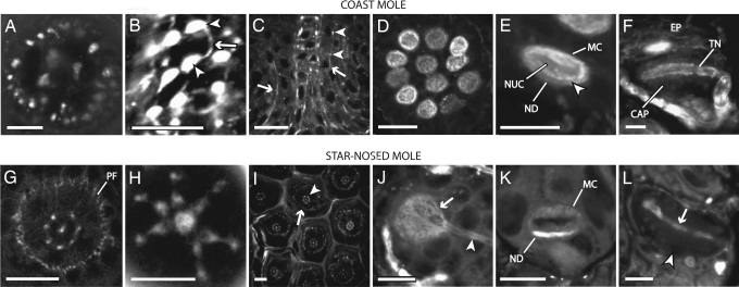
Fig. 3.
Confocal images showing AM1-43 labeling in Eimer’s organ of the coast mole (A–F) and star-nosed mole (G–L). (A) A horizontal section near the middle of the cell column showing a ring of 22 satellite nerve endings surrounding the central nerve fibers in the coast mole. (B) Brightly labeled varicosities (arrowheads) connected by thin neurites (arrow) give the free nerve ending terminals a bead-on-a-string appearance. (C) Vertical section in the coast mole showing short lengths of peripheral fibers on both sides of the central cell column fibers (arrows). The terminals are smaller than their central column counterparts but have a similar bead-on-a-string morphology (arrowheads). (D) Clustering of Merkel cell–neurite complexes at the base of the coast mole organ. (E) Magnified view of a Merkel cell–neurite complex (MC) with its characteristic lobulated nucleus (NUC) and terminal neurite disk (ND). The division is visible between the terminal neurite disk and the Merkel cell (arrowhead). (F) A vertical section through a lamellated corpuscle. The terminal neurite (TN) is aligned within the capsule (CAP) parallel to the skin surface. The whole complex sits directly below the epidermis (EP). (G) Horizontal section from a star-nosed mole ≈15 μm below the apex of the Eimer’s organ showing seven satellite fibers surrounding a single central fiber. The second ring of smaller diameter peripheral fibers is evident (PF) surrounding the center and satellite fibers. (H) Horizontal view of the most superficial free nerve ending terminals of an Eimer’s organ. The swellings of the satellite free nerve endings extend inward in close apposition to the central swelling. (I) Horizontal section showing star-nosed mole Eimer’s organs in a hexagonal array. The central free nerve endings (arrowhead) are surrounded by the peripheral ring of smaller, less organized nerve terminals (arrow). (J) Horizontal view showing the AM1-43-labeled neurite disk (arrow) and afferent neuron (arrowhead) of a single Merkel cell–neurite complex. (K) Vertical view of a Merkel cell–neurite complex. Note the higher level of staining of the neurite disk (ND) compared with the Merkel cell (MC). (L) Horizontal section showing a lamellated corpuscle. The terminal neurite (arrow) runs through the center of the capsule (arrowhead). (Scale bars: 10 μm in A, B, E, F, H, and J–L; 20 μm in C, D, G, and I.)

