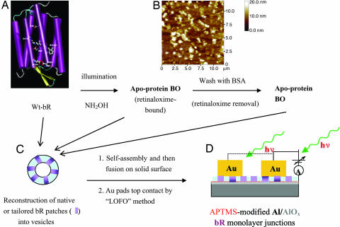
Fig. 1.
Scheme of bR chemical tailoring and of the metal–protein–metal junction preparation. (A) Schematic representation of the 3D structure of bR. The seven α-helical domains form a transmembrane pore. The retinylidene residue is linked to the protein moiety via a protonated Schiff base linkage to Lys-216. (B) Representative AFM image (12 × 12 μm) of native bR patches prepared by 5 min of adsorption on an Al/AlOx substrate derivatized with APTMS. (C) Schematic of bR-containing vesicle. (D) Schematic of a Au/(single-bR-layer)/(APTMS-on-AlOx)/Al junction and measuring scheme.

