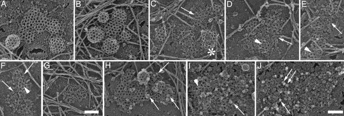
Fig. 4.
AP2 forms membrane microdomains in clathrin-depleted HeLa cells. Shown are views of the inner surface of unroofed HeLa cells that expressed their normal content of clathrin (A, B, and H), ≈40% clathrin (C–F), and <20% clathrin (G and I). Control cells (A, B, and H) are characterized by mainly hexagonal flat clathrin lattices and coated buds. Cells with reduced clathrin content reveal fewer buds (see asterisk in C) and incomplete lattices (e.g., arrow in C and arrowheads in D–F). Arrows in D–F point at clustered particles that are uncovered by the removal of clathrin. A cluster apparently devoid of clathrin is shown in G. They were identified as AP2 molecules by ImmunoGold labeling with AP.6 antibody followed by 10-nm gold-labeled anti-mouse IgG [arrows in control (H) and knockdown cells (I and J)]. The arrowhead in I denotes residual clathrin lattice structures. Anaglyph stereo views reveal that the AP2 membrane domains are almost completely flat (see Fig. 8). (Scale bars: 100 nm.)

