Figure 3.
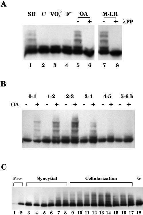
DAH is hyperphosphorylated during cellularization. (A) Different phosphatase inhibitors, VO43−, F−, okadaic acid (OA), and microcystin (M-LR), were included in the homogenization buffer to prepare embryonic extracts. Embryos were also lysed in SDS sample buffer directly (SB) or in homogenization buffer (C). Total proteins (100 μg) were loaded for each sample, and Western blotting with DAH antibody was performed. Several slow-migrating species were detected when OA and M-LR were added (lanes 5 and 7), and they all collapsed to the lowest band after the treatment of λ protein phosphatase (λPP; lanes 6 and 8). (B) Hourly staged embryos during the first 6 h of development were prepared with or without OA for Western blot analysis. DAH phosphorylation reached its peak at 2–3 h. (C) Single embryos were staged according to their morphology and nuclear density by light microscopy. Embryos in the preblastoderm (Pre-), syncytial blastoderm, cellularization, and gastrulation (G) stages were lysed in SDS sample buffer and loaded in the order of development. Embryos in lanes 9 and 10 had not extended their cleavage furrows. Embryos in lanes 11–14 were all in the slow phase of membrane invagination, followed by fast-phase embryos in lanes 15–17. DAH phosphorylation is most extensive during the slow phase of cellularization (lanes 11–14).

