Figure 6.
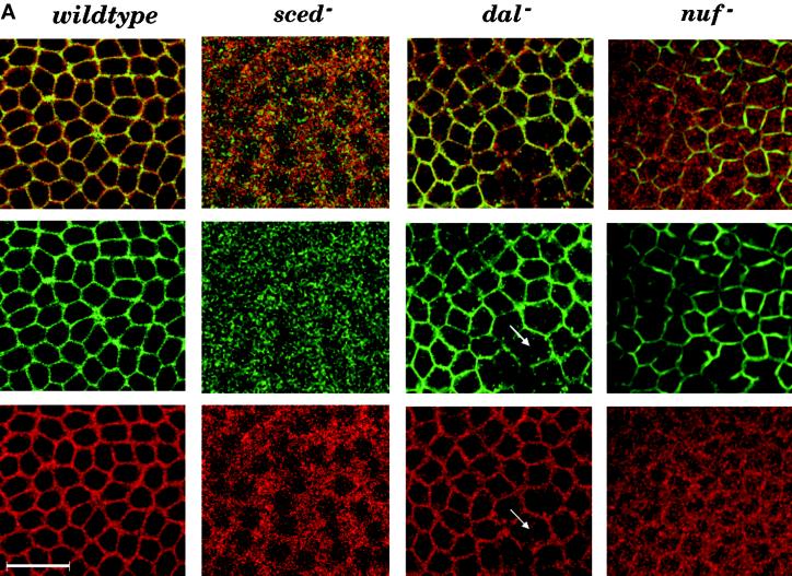
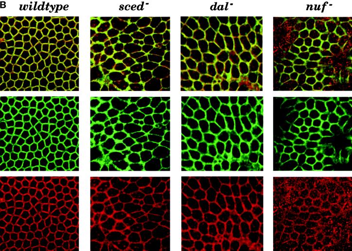
DAH mislocalizes in nuf mutants. (A) Wild-type and mutant embryos (sced−, dal−, and nuf−) in cycle 13 metaphase were double stained with fluorescein-conjugated phalloidin (green channel) and DAH antibody followed by a Cy3-conjugated secondary antibody (red channel). The top panels are the merged images of the green and red channels below them. The arrows point to the disrupted actin network in the dal mutant panels. DAH colocalizes with actin in the wild-type embryo (left columns) and in the sced and dal mutants (middle columns). However, DAH staining is largely diffused in the nuf mutant, although incomplete actin hexagons are present (right columns). Bar, 25 μm. (B) Wild-type and mutant embryos in cellularization were prepared as in A. In the wild-type embryo, DAH concentrates in the cleavage furrows (left columns). It localizes to the cleavage furrows in the sced and dal mutants (middle columns), whereas the DAH localization in nuf remains defective (right columns).


