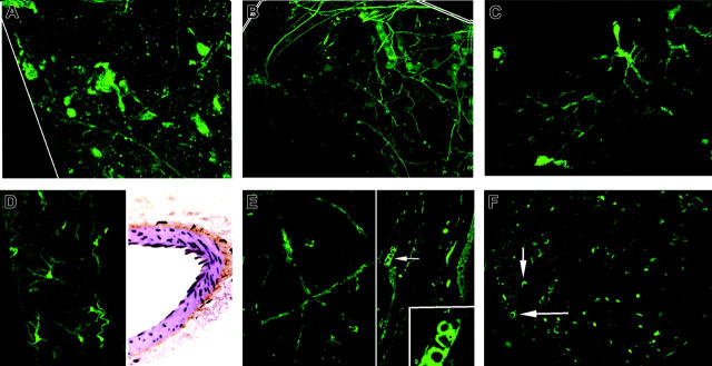
Figure 2.
Vessel-wall t-PA/EGFP expression. (A) Aorta surface: 3D Z-axis composite of adventitial layers shows positve expression by a few discrete axons compatible with sparse innervation. (B) Carotid artery surface: 3D Z-axis composite of adventitial layers reveals positive expression by numerous postitve axons. (C) Renal artery surface: positive expression is confined to a fairly dense nerve net with prominent varicose dilations. (D) Mesenteric artery surface: numerous axons with bright terminal varicosities appear in deep adventitial layer (left). Section immunostained for t-PA shows dense subadventitial plexus on outer smooth-muscle surface (brown color; right). (E) Ear skin surface: positive expression appears confined to walls of branching arteriole, absent from numerous adjacent venules and capillaries. (F) Sciatic nerve section: expression is positive in walls of multiple nutrient vasa nervosa (arrows) and in scattered cross-cut axons. Magnifications are ×40, except as noted.

