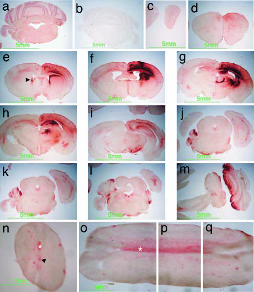
Fig. 1.
β-hexosaminidase activity in murine brain and spinal cord after gene therapy. Red staining indicates β-hexosaminidase product in wild-type control (a) and in rAAV2/2β-transduced Sandhoff mice (c–q), absent in untreated Sandhoff mice (b). rAAV2/2β was stereotaxically inoculated at a single site in the right striatum at 4 weeks and killed at the humane end point of 35 weeks of age. Staining extends from the olfactory bulbs (c) to the spinal cord (n–q). Staining is also observed in ependymal cells in lateral ventricles and the central canal of the spinal cord (arrowheads in e and n, respectively) and in white matter, particularly evident in the pyramidal tract (asterisk in n–q). Reconstruction of a single longitudinal section of spinal cord is depicted in o–q. Sections c–q were obtained from the animal shown in Movie 2, which is published as supporting information on the PNAS web site.

