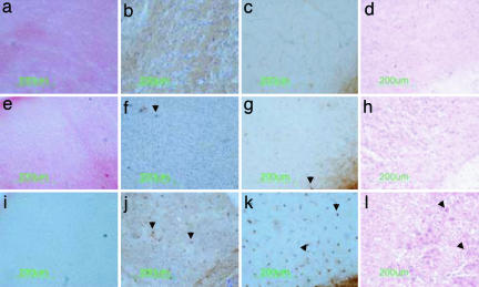
Fig. 3.
Relationship between β-hexosaminidase activity, glycosphingolipid storage, and inflammatory cells in the cerebral cortex. Coronal sections from wild type aged 16 weeks (a–d), rAAV2/2α+β-transduced aged 29 weeks (humane end point) (e–h), and untransduced Sandhoff mice aged 17 weeks (i–l) were prepared consecutively. Virus was injected at 4 weeks of age. The β-hexosaminidase reaction product stains red (a and e) and is absent in untransduced Sandhoff mice (i).Glycospingolipid storage, detected by neuronal PAS staining, occurs particularly in layers IV and V of the cerebral cortex of untreated Sandhoff mice (arrowheads in l) but was undetectable in cortex from wild-type (d) or transduced Sandhoff mice (h). Activated microglia/macrophages were recognized by immunostaining of the cell-specific marker, CD68 (b, f, and j), and by binding to isolectin B4 (c, g, and k). No cells of microglia/macrophage lineage were detected in wild-type cortex (b and c), and only a few were seen in transduced Sandhoff mice (arrowheads in f and g). Cerebral cortex from untransduced Sandhoff mice contained numerous activated microglia and macrophages (arrowheads in j and k). The number of neurons staining with PAS and the presence of cells recognized by G. simplicifolia isolectin B4 (GSIB4) and CD68 antibodies inversely depended on enzymatic activity.

