
Fig. 1.
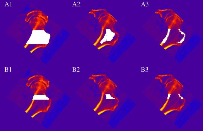
vQCT regions of interest in the proximal femur. Regions of interest are white pixels superimposed on image data. vQCT images showing (1) iBMD (2) tBMD and (3) cBMD in the (A) overall proximal femur and in (B) the FN. The TR is the region outside the FN but within the overall proximal femoral region.