
Figure 2. Immunofluorescence analysis of cyst phenotype and laminin α5 expression.
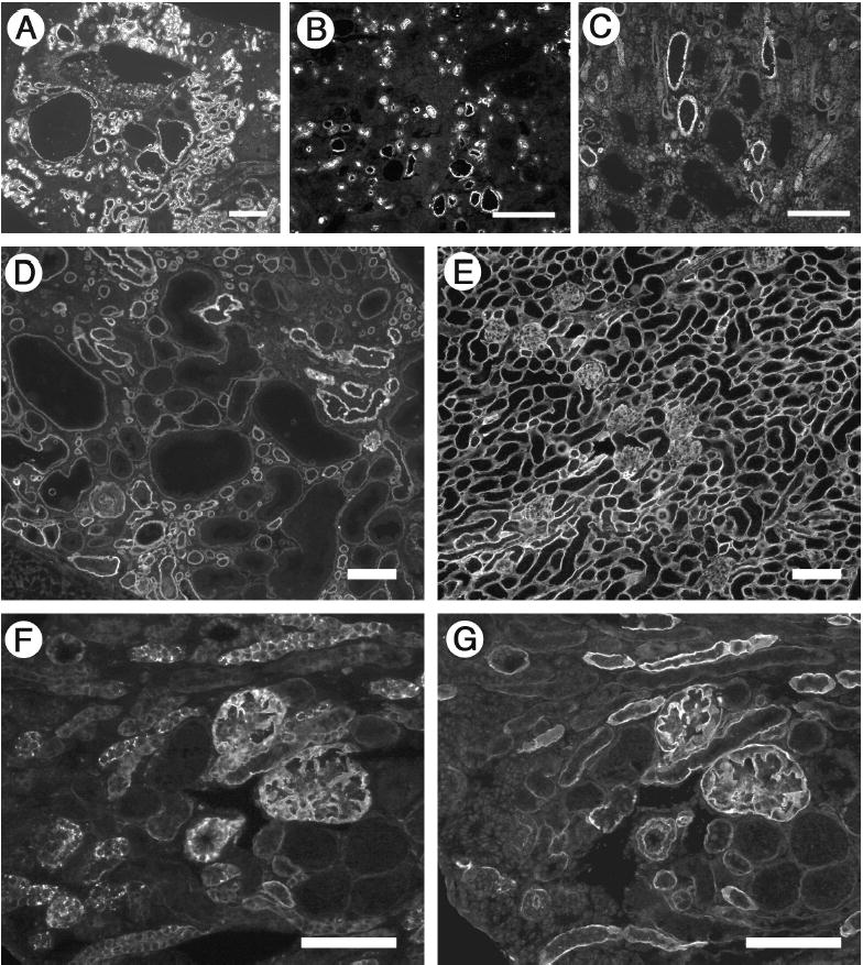
(A–C) Cysts derive from multiple tubular segments. Frozen sections of P23 cystic kidneys were stained with FITC-conjugated Lotus tetragonolobus lectin to label proximal tubules (A), antibody to Tamm-Horsfall protein to label thick ascending limb of the loop of Henle (B), and antibody to cytokeratin 8 to label collecting ducts (C). Consistent with a de-differentiated phenotype, some large cysts failed to label with any of the markers. (D and E) Laminin α5 levels were reduced in Lama5neo/neo kidney (D) compared to control (E) at P21. Laminin α5 deposition was uniform in the control but heterogeneous in the mutant; most cysts showed decreased to nearly undetectable levels of α5. (F and G) Truncated laminin α5 accumulates inside cells. Serial sections from a Lama5neo/neo kidney were stained with antibodies whose epitopes are either NH2-terminal (F) or COOH-terminal (G) of the truncation in domain LEa. Scale bars in A–C: 200 μm; in D-G: 100 μm.