
Figure 4. Proliferation and apoptosis assays.
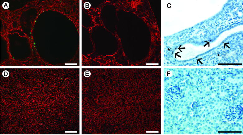
(A and D) Assay for BrdU (green), counterstained with anti-laminin-111 to label basement membranes (red), shows proliferating cells in cyst-lining epithelia (A). BrdU-positive cells were also occasionally observed in the interstitium and in normal tubules in mutants (A) but were rarely found in control (D). (B and E) Serial sections with respect to A and D show staining for laminin α5, which is focally decreased in cysts (B) but more homogenously expressed in control (E). (C and F) TUNEL staining at P20. Apoptotic cells were present in cyst-lining epithelia (arrows in C), as well as interstitium (asterisks), but were absent in controls (F). Scale bars: 100 μm.