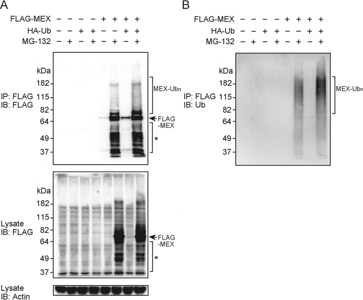
Figure 3. Ubiquitination of MEX in vivo.
(A) HEK-293T cells were co-transfected with HA-tagged ubiquitin (HA–Ub) and/or FLAG-tagged MEX (FLAG–MEX). Cells were treated with 10 μM MG-132 or left untreated for 24 h. The lysates were subjected to immunoprecipitation (IP) with an anti-FLAG Ab, and immunocomplexes were eluted with excess FLAG peptide. The eluted proteins were analysed by immunoblotting (IB) with an anti-FLAG Ab (top panel). Total lysates were blotted with an anti-FLAG Ab (middle panel) or an anti-actin Ab (bottom panel). (B) After stripping, the membrane used in (A) was immunoblotted with an anti-Ub Ab. Ubiquitinated full-length, and degraded (*) MEX proteins are indicated by brackets.

