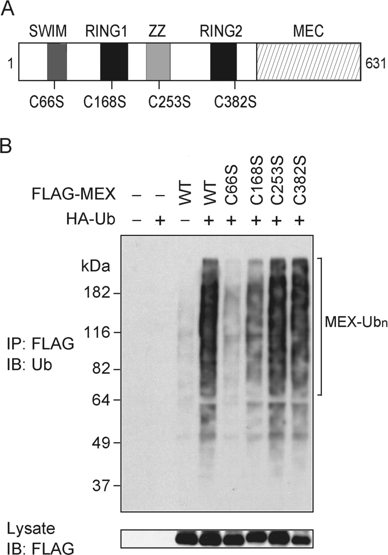
Figure 5. Mutational analysis of MEX ubiquitination.
(A) Schematic diagram of mutant MEX proteins. The positions of point mutations introduced into the MEX gene are shown. (B) HEK-293T cells were co-transfected with HA-tagged Ub and/or the indicated FLAG-tagged MEX constructs. Cells were treated as described in Figure 3(A). Immunocomplexes were analysed by immunoblotting (IB) with an anti-Ub Ab (upper panel). Total lysates were blotted with an anti-FLAG Ab (lower panel). IP, immunoprecipitate.

