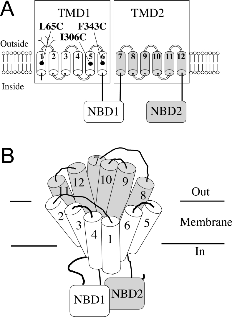
Figure 1. Schematic models of P-gp.
(A) The 12 TMs of P-gp are shown as numbered cylinders. The branched lines represent glycosylation sites. The locations of residues L65C in TM1, I306C in TM5 and F343C in TM6 are shown. The ATPase activities of these mutants were permanently activated after modification with thiol-reactive drug substrate analogues. (B) Predicted packing of the TM segments of P-gp. The common drug-binding pocket is at the interface between TMD1 and TMD2.

