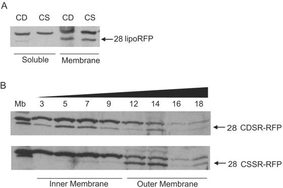
FIG. 2.
Localization of lipoRFPs CDSR-RFP and CSSR-RFP. (A) Analysis of membrane and soluble protein fractions. Samples were derived from identical starting cell suspension volumes. (B) Membranes were separated by flotation sucrose density gradient centrifugation. Total membranes were used as a control (Mb), and the sucrose gradient fraction number is indicated above. The black triangle indicates the increasing concentration of sucrose in the gradient fractions. Proteins in samples were separated by SDS-PAGE and immunoblotted with antibodies against mRFP1. The identities and molecular sizes (kilodaltons) of the lipoRFPs are indicated.

