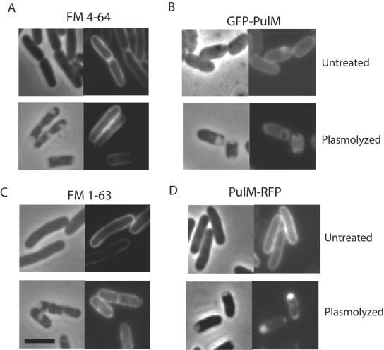
FIG. 3.
Outer membrane staining of lipophilic FM dyes and inner membrane localization of PulM. Live cells were left untreated or plasmolyzed in the presence of 5 μg/ml FM 4-64 (A) or FM 1-63 (C). Strains producing GFP-PulM (B) or PulM-RFP (D) were left untreated or plasmolyzed. Each panel consists of a phase-contrast image (left) and the corresponding fluorescence image. The black bar is 3 μm in length.

