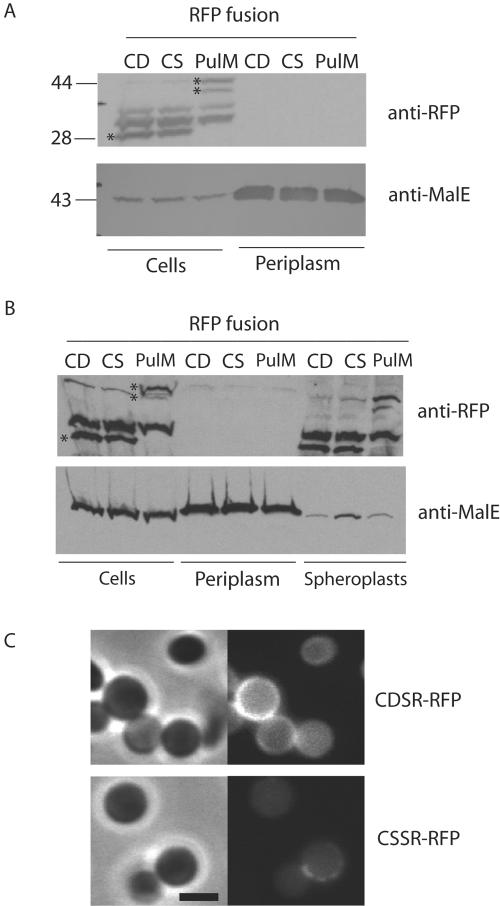
FIG. 4.
Plasmolyzed cells do not release fluorescent lipoproteins into the periplasm. (A) Proteins released by osmotic shock of cells producing CDSR-RFP, CSSR-RFP, or PulM-RFP after plasmolysis with 15% sucrose. (B) Proteins released by converting cells to spheroplasts by digesting the peptidoglycan layer with lysozyme following sucrose plasmolysis. In panels A and B, the samples were derived from the same volume of initial cell suspension. Proteins were separated by SDS-PAGE and immunoblotted with antibodies against mRFP1 or MalE. Asterisks indicate the lipoRFPs and PulM-RFP, and their molecular sizes (kilodaltons) are shown at the left of panel A. (C) Spheroplasts of cells producing CDSR-RFP and CSSR-RFP examined by phase-contrast and fluorescence microscopy. The black bar is 2 μm in length.

