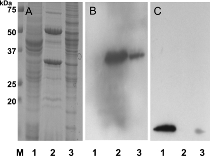
FIG. 2.
Subcellular localization of TcdC in C. difficile cells. (A) Analysis by SDS-PAGE of membrane and cytosolic proteins harvested from C. difficile strain VPI 10463 grown in tryptone-yeast extract-glucose medium for 4 h. Lanes: 1, cytosolic proteins; 2, Triton X-100-soluble membrane proteins; 3, Triton X-100-insoluble proteins. The proteins were stained with Coomassie brilliant blue. (B) Immunodetection of TcdC by using anti-TcdC antibody. (C) Immunodetection of L7/L12 ribosomal protein using monoclonal anti-L7/L12 streptococcal ribosomal proteins (dilution, 1:1,000). The protein samples in panels B and C correspond to those in the same-numbered lanes in panel A.

