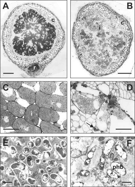
FIG. 4.
Structures of Vigna unguiculata nodules formed by Rhizobium sp. strain NGR234 (A, C, and E) and the y4gM mutant NGRΩgM (B, D, and F). (A and B) Low-magnification light micrographs of whole nodules. Magnification, ×35. Bar, 200 μm. (C and D) High-magnification light micrographs of nodule sections. Magnification, ×312. Bar, 32 μm. (E and F) Electron micrographs of bacteroidal tissue. Magnification, ×8,650. Bar, 2 μm. Abbreviations: b, bacteroids; c, nodule cortex; i, infected cells; phb, polyhydroxybutyrate; r, roots. The black arrow points to an amyloplast containing starch granules, and the black circle indicates peribacteroid membranes.

