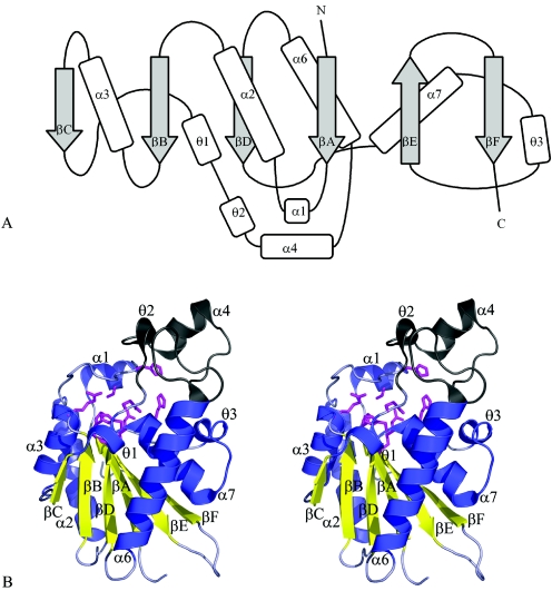
FIG. 1.
Structure of Rv3214 from M. tuberculosis. (A) Topology diagram. Secondary structures, labeled to follow the nomenclature of Rigden et al. (38), comprise the following residues: βA, 7 to 13; α1, 16 to 22; α2, 33 to 50; βB, 55 to 60; α3, 62 to 73; βC, 77 to 80; θ1, 80 to 84; θ2, 88 to 92; α4, 95 to 104; α6, 118 to 137; βD, 141 to 146; α7, 147 to 160; θ3, 162 to 166; βE, 174 to 180; and βF, 187 to 193. (B) Stereoview of the Rv3214 monomer. The major parts of the molecule are shown in blue (helices) and gold (β-strands), with the small subdomain shown in gray and residues that contribute to the active site shown in magenta, in stick mode.

