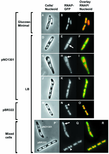
FIG. 2.
Cell morphology and location and distribution of RNA polymerase in cells carrying the plasmid-borne rrnB operon in pNO1301 grown in rich and minimal media. (A, D, G, J, M, and P) Merged phase-contrast and DAPI fluorescence images. (B, E, H, K, N, and Q) Fluorescence from the RNAP-GFP fusion protein. (C, F, I, L, O, and R) Overlays of the fluorescence from the RNAP-GFP fusion protein (green) and the fluorescence of the DAPI-stained nucleoid (red). (P, Q, and R) Comparison of cell morphology and location and distribution of RNA polymerase in cells carrying either pNO1301 or pBR322 in the same microscope field. Cells carrying pNO1301 and pBR322 were grown in LB medium and mixed at a ratio of 1:1 before fixation, and this was followed by fluorescence microscopy. Representative images are shown. Note that while transcription foci are evident in the nucleoids of cells harboring pBR322 (arrowhead), they are diminished in the nucleoids of cells harboring pNO1301. Although most of the RNAP-GFP fluorescence signals in the cytoplasm are diffuse, sometimes concentrated signals are apparent in the cytoplasm of cells harboring pNO1301 (arrows). For simplicity, only one representative focus is indicated.

