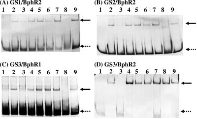
FIG. 5.
Gel shift assays of the BphR1 and BphR2 proteins.The 3′-end-labeled GS1 (A), GS2 (B), and GS3 (C and D) DNA fragments containing the possible operator regions of bphR1, bphA1, and bphR2-salA were incubated without protein (lane 1) or with purified BphR1 or BphR2 protein (lanes 2 to 9). The amount of the BphR2 protein (A, B, and D) used was 100 ng (lanes 2 to 9). The amount of the BphR1 protein (C) used was 2 μg (lanes 2 to 9). Excess unlabeled DNA (lane 3) or unrelated DNA (lane 4) was added. Biphenyl (lane 5), HOPD (lane 6), salicylate (lane 7), HMSA (lane 8), or benzoate (lane 9) was added at a concentration of 1 mM. The dotted arrows indicate free DNA bands, and the solid arrows indicate retarded DNA bands.

