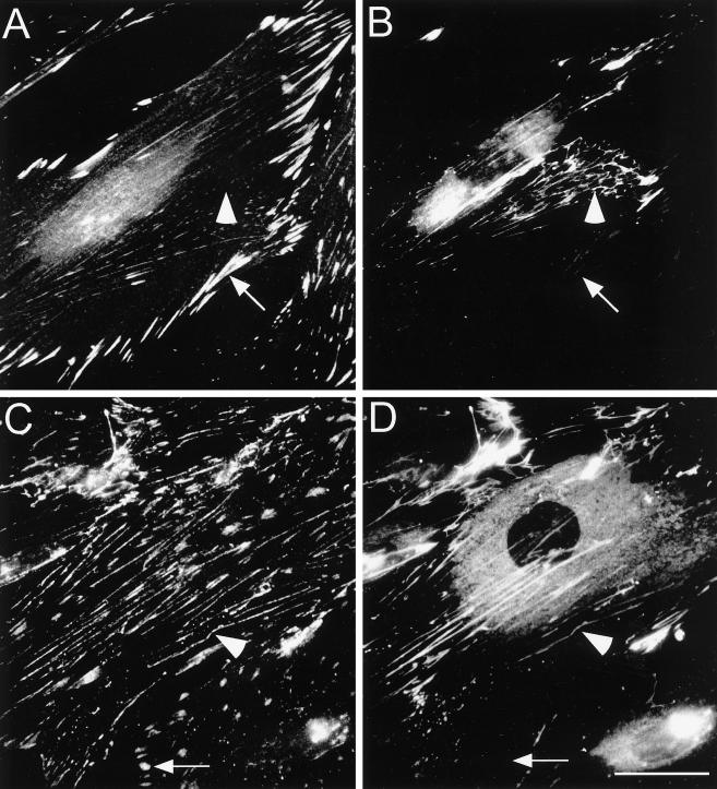
Figure 1.
Distribution of fibronectin, paxillin and tensin in primary human fibroblasts. Cells were cultured for 16 h on coverslips. Double immunofluorescence staining was then performed with antibodies to paxillin (A) and fibronectin (B) or with tensin (C) and fibronectin (D). Note the localization of paxillin and tensin in FCs (arrows) and tensin along fibronectin fibrils (arrowheads). Bar, 20 μm.

