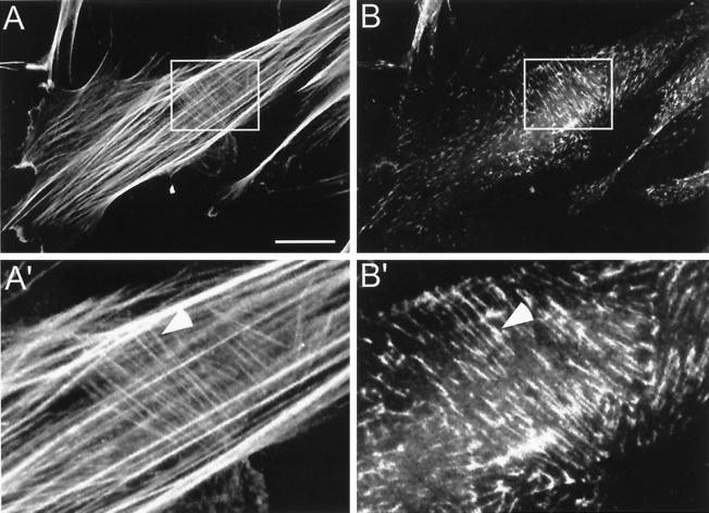
Figure 3.
F-actin localization at cell-matrix adhesions. Primary human fibroblasts were cultured on coverslips for 16 h. Double immunofluorescence staining was then performed for (A) F-actin (rhodamine-conjugated phalloidin) and (B) α5 integrin (rat anti-α5 integrin monoclonal antibody followed by FITC-conjugated goat anti-rat antibody). (A′) and (B′) High-power magnification of the regions defined by rectangles in (A) and (B), respectively. Arrowheads indicate the location of fibrillar adhesions and thin actin filaments. Bar, 15 μm.

