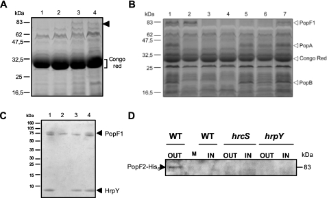
FIG. 1.
Secretion of PopF1 and PopF2 and detection of PopF1 in Hrp pili preparations. (A) Protein secretion after growth of various R. solanacearum strains carrying pAM5 in the presence of Congo red. Proteins present in CSP-CRs prepared from (lane 1) hrp mutant strain GMI1410(hrpY)/pAM5, (lane 2) hrp mutant strain GMI1402(hrcS)/pAM5, (lane 3) GMI1551(popA)/pAM5, and (lane 4) GMI1000/pAM5 grown in MMG supplemented with Congo red were separated by SDS-PAGE and visualized by Coomassie blue staining. The arrowhead shows the new putative 80-kDa Hrp-secreted protein. The bracket indicates the position of Congo red, which in SDS-PAGE exhibits the same electrophoretic mobility as a 28-kDa protein. (B) Proteins present in CSP-CRs prepared from GMI1000/pAM5 (lane 1), GMI1551(popA)/pAM5 (lane 2), hrp mutant strain GMI1402(hrcS)/pAM5 (lane 3), GMI1410 (hrpY)/pAM5 (lane 4), GMI1663 (popF1)/pAM5 (lane 5), GMI1666 (popF1 popF2)/pAM5 (lane 6), and GMI1664 (popF2)/pAM5 (lane 7), grown in MMG supplemented with Congo red, were separated by SDS-PAGE and visualized by silver staining. Positions of PopA, PopB, PopF1, and Congo red are shown by arrowheads. (C) Characterization of PopF1 in Hrp pili preparations. Shown is a Tris-Tricine SDS-PAGE electrophoresis gel (10 to 20% acrylamide) loaded with protein samples prepared from the surface of the following strains grown on MMG: (lane 1) GMI1000, (lane 2) GMI1584(hrcV) mutant, (lane 3) GMI1663(popF1) mutant, and (lane 4) GMI1664(popF2) mutant. Proteins were revealed by Coomassie blue staining. Arrowheads show PopF1 and HrpY. (D) Secretion of PopF2-His6 in wild-type (WT) or hrp background. Bacterial strains GMI1668(popF2-His6), GMI1669(hrcS popF2-His6), and GMI1670(hrpY popF2-His6) were cultivated in MMG supplemented with Congo red. Concentrated supernatant preparations (CSP-CR) (OUT) and concentrated lysate preparations (CLP-CR) (IN) were prepared from these cultures, and their protein contents were separated by SDS-PAGE and visualized by immunoblotting with an anti-His6 monoclonal antibody. M, size marker.

