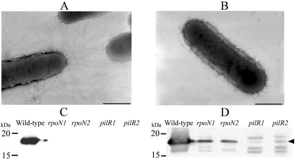
FIG. 2.
Absence of fimbriae in pilR and rpoN mutants. The presence of surface fimbriae was observed using electron microscopy. (A and B) Wild-type cells showing the presence of fimbriae (A) and the pilR mutant lacking surface fimbriae (B). Note that the rpoN mutants appeared similar to those shown in B. Bars equal 1 μm. The presence of surface fimbriae was also determined using Western immunoblotting with fimbrial antisera on sheared fimbriae (C) and whole-cell lysates (D).

