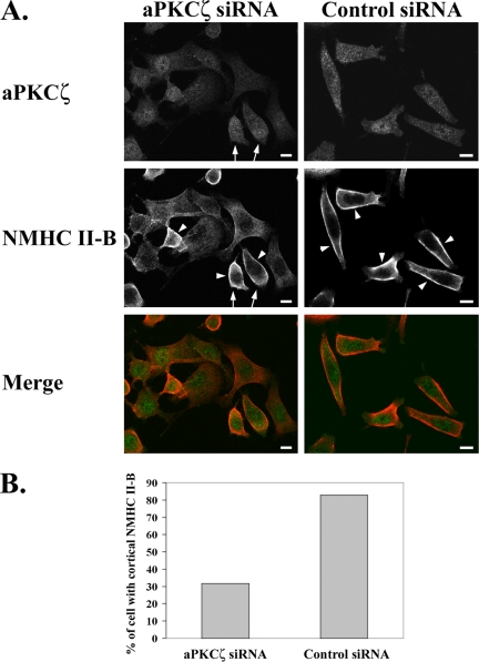
Figure 10.
Reduced aPKCζ expression alters EGF-dependent NMHC II-B cellular localization and organization. (A) immunofluorescence staining of aPKCζ (top panels, green) and NMHC II-B (middle panels, red) in TSU-pr1 cells transfected with either aPKCζ siRNA (left panels) or control siRNA (right panels) and stimulated with EGF for 1 min. Arrows point to cells that show relatively high levels of aPKCζ expression in the aPKCζ siRNA cell population. Arrowheads point to cortical localization of NMHC II-B. Bar, 10 μm. (B) Quantification of the number of cells that exhibit cortical localization of NMHC II-B in cells transfected with aPKCζ siRNA compared with control siRNA. The results are presented as the percentage of cells that show cortical localization out of the total number of cells counted in 10 random fields (average total of cells counted for each type of siRNA treatment: n = 200).

