Figure 9.
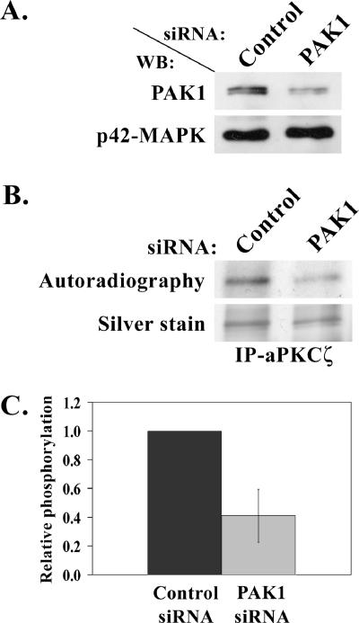
PAK1 is involved in aPKCζ phosphorylation in vivo. (A) A representative Western blot showing PAK1 protein level after transfection of TSU-pr1 cells with PAK1 siRNA (PAK1) or fluorescein-conjugated control siRNA (Control). Cells were transfected with the siRNA 48 h before the experiment. To determine the expression level of PAK1 in siRNA cells, 10% of the lysates were subjected to Western blot analysis using PAK1 antibody followed by p42-MAPK antibody, as a loading control. (B) A representative in vivo phosphorylation experiment of aPKCζ. aPKCζ was immunoprecipitated from cell lysates transfected as described in A. The immunoprecipitates were separated on SDS-PAGE, which was then stained with silver stain. The gel was scanned, dried, and exposed to film (Autoradiography). (C) A plot representing the amount of phosphorylated aPKCζ in cells treated with PAK1 siRNA relative to the amount of phosphorylated aPKCζ in the control cells. The amount of phosphorylated aPKCζ was calculated by dividing the value of the phosphorylated aPKCζ band by the value of the total aPKCζ band from the gel. The plot summarizes results of three experiments such as the one described in B.

