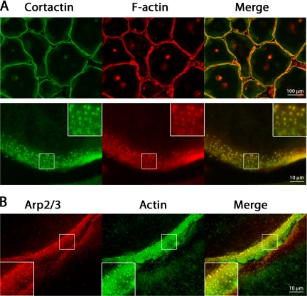
Figure 4.
Cortactin, actin, and Arp2/3 complex localization to podosomes. Osteoclasts on glass were costained for cortactin and F-actin, or Arp3 and actin. (A) Low- and high-magnification fluorescence images of osteoclasts stained with anti-cortactin antibody and rhodamine-phalloidin. (B) Higher magnification images of the periphery of two adjacent osteoclasts stained with anti-Arp3 and anti-actin antibodies. In all images, podosomes look like punctate, fluorescent structures. Diffuse staining for actin, cortactin, and Arp3 is observed between podosomes.

