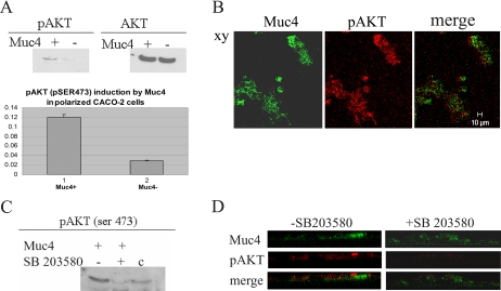
Figure 11.
Analysis of the induction of Akt phosphorylation at serine 473 in Muc4-expressing CACO-2 cells via p38 activation. (A) Immunoblot analyses of the phosphorylated and total forms of Akt of CACO-2 cell lysates in Muc4-transfected cells. Plot of the induction of phosphorylated Akt at serine 473 was based on the immunoblot data. Control cells were transfected with empty vector and analyzed in the same manner. (B) Confocal immunofluorescence localization of pAkt in a confluent monolayer of CACO-2 cells. First panel, Muc4-expressing cells (green); second panel, staining with monoclonal anti-pAkt on serine 473 shows apical staining for cells (red). Note that only cells expressing Muc4 have activated Akt. Confocal sections are shown with the apical side up, x-y plane; bar 10 μm. (C) Immunoblot analyses of the phosphorylated Akt at serine 473 of CACO-2 cell lysates of Muc4-transfected cells treated with or without p38 inhibitor SB203580. A375 cell lysates were used as control. (D) X-z plane by confocal immunofluorescence of pAkt in a confluent monolayer of CACO-2 cells in the presence and in the absence of SB203580. First panel, Muc4 (green); second panel, staining with polyclonal anti-pAkt; and third panel, merge of panels one and two.

