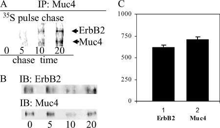
Figure 3.
Time of formation and stoichiometry of the Muc4–ErbB2 complex in A375 cells by 35S labeling. (A) Tetracycline-controlled expression of Muc4 in A375 cells was initiated by the removal of tetracycline, and the cells were pulse-labeled 10 min with 35S amino acids and chased for periods of 0–20 min. (B) Muc4–ErbB2 complex was immunoprecipitated with anti-Muc4. The presence and location of ErbB2 on the fluorography were confirmed by immunoblot using mAb Neomarkers 10. The presence and location of Muc4 were confirmed by immunoblot (bottom left panel) after stripping of the membrane. (C) Graph of the relative intensities of the bands detected by the 35S fluorography after subtraction of the background and normalization by the ratio of the content of cysteines and methionines in both molecules. This graph indicates a stoichiometry of 1:1 for the Muc4–ErbB2 complex.

