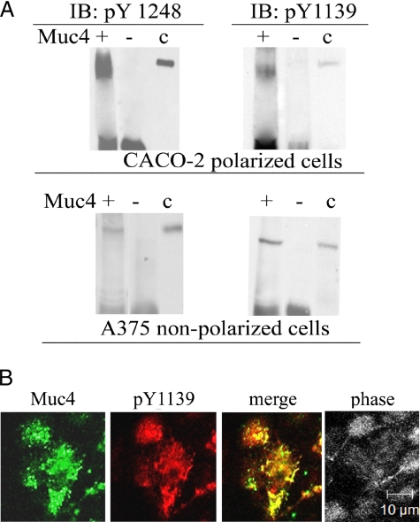
Figure 6.
Phosphorylation of ErbB2 in the Muc4–ErbB2 complex in CACO-2 and A375 cells. (A) Lysates from Muc4-transfected CACO-2 and A375 cells were immunoprecipitated with anti-Muc4. The precipitates were then immunoblotted using antibodies against activated ErbB2, phosphorylated on tyrosines 1248 and 1139, showing that ErbB2 from both cell types was phosphorylated on both sites. (B) Confocal immunofluorescence showing colocalization of ErbB2 activated at tyrosine 1139 with polyclonal anti-ErbB2 pY1139 and Muc4 in a confluent monolayer of CACO-2 cells. Confocal sections are shown with the apical side up, x-y plane; bar, 10 μm.

