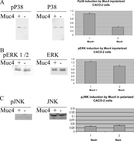
Figure 8.
Analysis of phosphorylation of p38, ERK, and JNK induced by Muc4 expression in polarized CACO-2 cells. (A) Immunoblot analyses of the phosphorylated and total forms of p38 of CACO-2 cell lysates of Muc4-transfected cells. Plot of the induction of phosphorylated p38 was based on the immunoblot data. Control cells were transfected with empty vector and analyzed in the same manner. (B) Immunoblot analyses of the phosphorylated and total forms of ERK 1/2 of CACO-2 cell lysates of Muc4-transfected cells. Plot of the increase in activated ERK levels was based on the immunoblot data. Control cells were transfected with empty vector and analyzed in the same manner. (C) Immunoblot analyses of the phosphorylated and total forms of JNK of CACO-2 cell lysates of Muc4-transfected cells. Plot of the activation of JNK was based on the immunoblot data. Control cells were transfected with empty vector and analyzed in the same manner.

