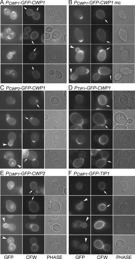
Figure 2.
Localization of GFP-Cwp1p to the birth scar and its dependence on both promoter sequences and sequences within or downstream of the coding region. GFP-tagged proteins were expressed from low-copy (A and C–F) or high-copy (B) plasmids in strain FY833. For each construct, >200 cells showing wall fluorescence were observed, and representative cells from four successive phases of the cell cycle are shown. (A and B) GFP-Cwp1p expressed from its own promoter (plasmids pGS-GFP-CWP1-low and pAR213). (C) GFP-Cwp1p expressed from the CWP2 promoter (plasmid pCWP2-GFP-CWP1). (D) GFP-Cwp1p expressed from the TIP1 promoter (plasmid pTIP1-GFP-CWP1). (E) GFP-Tip1p expressed from the CWP1 promoter (plasmid pCWP1-GFP-TIP1). (F) GFP-Cwp2p expressed from the CWP1 promoter (plasmid pCWP1-GFP-CWP2). Phase, phase-contrast images; arrows, birth scars; arrowheads, buds with strong fluorescence.

