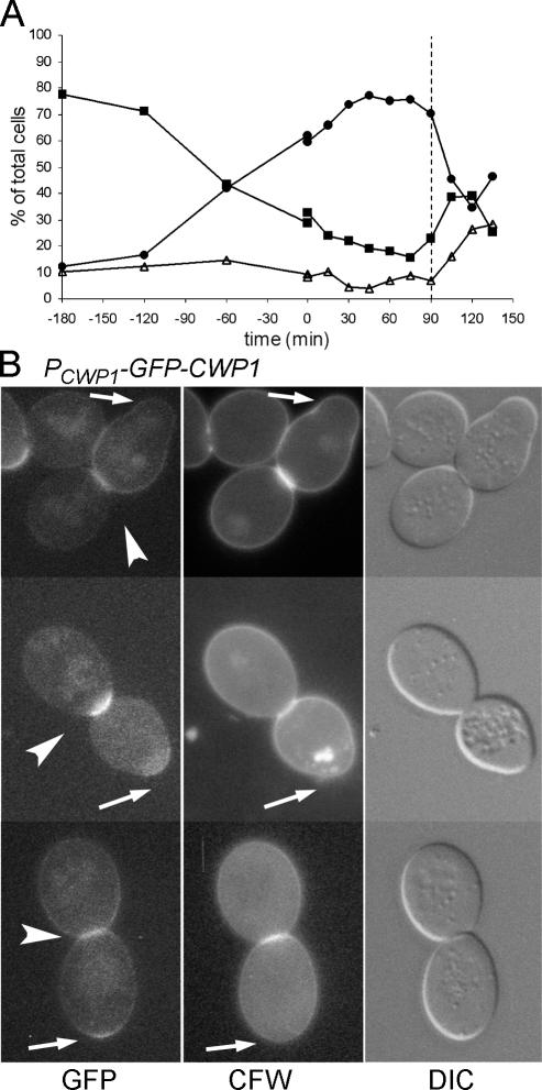
Figure 4.
Incorporation of GFP-Cwp1p into the secondary septum of the daughter cell after cytokinesis. Cells of strain LSY265 containing plasmid pGS-GFP-CWP1-low were synchronized by adding hydroxyurea at t = −180 min and washing it out at t = 0 (see Materials and Methods). (A) Synchronization shown as percentages of unbudded (■), small-budded (▵), and large-budded (●) cells. (B) Fluorescence and DIC images of large-budded cells from the 90-min sample (A, dashed line). Arrows, birth scars of the mother cells; arrowheads, GFP-Cwp1p on the daughter side of the necks.

