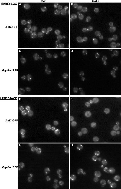
Figure 8.
AP-1 is mislocalized in laa1Δ cells. Wild-type (A, C, E, and G) and laa1Δ (B, D, F, and H) cells expressing GFP-tagged β1 (Apl2-GFP; A and B and E and F) or mRFP-tagged Gga2p (Gga2-mRFP; C and D and G and H) were grown to a late stage (E–H) and visualized by epifluorescence microscopy. The late stage cultures were diluted and grown to early log phase before visualizing again (A–D).

