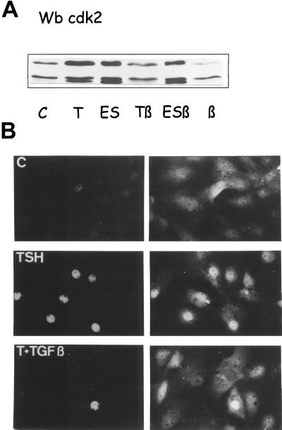
Figure 4.
Inhibition by TGFβ of the TSH-stimulated phosphorylation (A) and nuclear translocation (B) of cdk2. Four-day-old dog thyrocytes were stimulated by TSH (1 mU/ml) or EGF (25 ng/ml) + serum (10%) (ES) alone or in combination with TGFβ1 (2 ng/ml) (β). (A) Activating Thr160 phosphorylation of cdk2 reflected by its downward electrophoretic shift (Gu et al., 1992) demonstrated by Western blotting from cells stimulated for 32 h. (B) Double immunofluorescence labeling of PCNA used as a cell cycle marker (left panels) and cdk2 (right panels) 26 h after cell stimulation. Notice the increased nuclear labeling of cdk2 in PCNA-positive cells observed in many TSH-stimulated cells but few cells treated with TSH+TGFβ.

