
Fig. 7. Model for regulation of CK1 activity by activation of group I mGluRs.
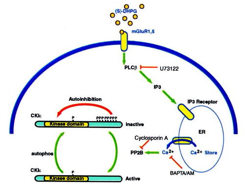
DHPG activates group I mGluRs that are coupled to PLCβ via Gq. Activation of PLCβ generates IP3, and IP3 binds to IP3 receptors on the endoplasmic reticulum and releases Ca2+ into the cytosol. Elevated intracellular Ca2+actives the Ca2+-dependent phosphatase calcineurin, which in turn dephosphorylates the regulatory autophosphorylation sites on CKIε. CKIε is transiently activated, but gradual autophosphorylation restores the inhibited level of kinase activity. A site that is basally phosphorylated is likely to be present within the kinase domain but does not appear to regulate enzyme activity.