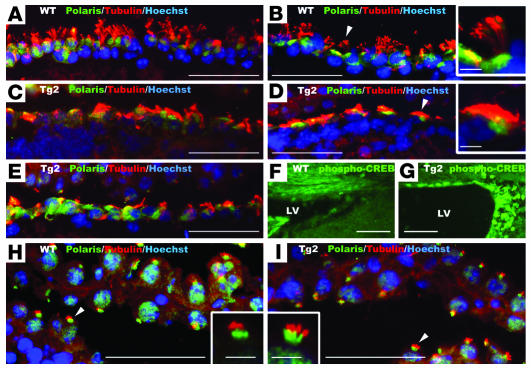
Figure 10. Disorganized cilia in ventricular ependyma but not in choroid plexus of Tg mice.
Brains from three 5-week-old Tg2 mice (C–E, G, and I) and 2 WT (A, B, F, and H) littermates were coronally sectioned. Sections were costained with Hoechst (blue), anti–acetylated α-tubulin (red), and anti-Polaris (green) (A–E, H, and I) or stained with anti–phospho-CREB (F and G). Insets in B, D, H, and I are magnified views of cilia (arrowheads) in the respective panels. Note that ependyma of Tg mice exhibited disorganized cilia and increased phospho-CREB reactivity. Scale bars: 50 μm in A–I and 5 μm in insets of B, D, H, and I.

