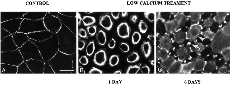
Figure 1.
Desmosomes of MDCK cells can be Ca-Dep or Ca-Ind. (A) Desmosomes of MDCK cells in confluent culture stained with mAb 11-5F to desmoplakin. Note that the desmosomes are located at the cell peripheries and that the staining is generally punctate. (B) A monolayer that has been cultured at confluent density for 24 h in SM and then treated with LCM-EGTA for 90 min, showing loss of intercellular contact and of desmosomal staining from the cell peripheries. This is indicative of Ca-Dep desmosomes. (C) A 6-d-confluent monolayer treated with LCM-EGTA for 90 min, showing partial loss of intercellular contact but persistence of joining processes with intense desmosomal staining (e.g., arrow). This is indicative of Ca-Ind desmosomes. Note that the cells in C show some cytoplasmic staining (e.g., arrowhead), probably indicative of the persistence of some Ca-Dep desmosomes. Bar, 20 μm.

