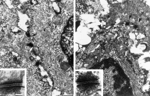
Figure 3.
Desmosomes of epithelial cell sheets in vivo are generally Ca-Ind. Electron micrographs of mouse esophageal epithelium that has been incubated for 6 h at 37°C in SM (A) or LCM-EGTA (B). Note that the intercellular spaces in B are generally wider than those in A but that the desmosomes (arrowheads) are still intact. The insets show higher-magnification images of individual desmosomes showing that those incubated in LCM-EGTA retain typical desmosomal ultrastructure. Bar, 1 μm; bar in inset, 0.1 μm.

