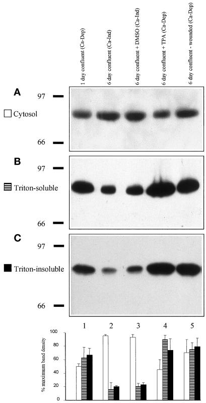
Figure 7.
Increase of PKCα in the Triton X-100–soluble and Triton X-100–insoluble fractions correlates with Ca-Dep of desmosomes. Immunoblotting for PKCα of cytosolic (A), Triton X-100–soluble (B), and Triton X-100–insoluble (C) fractions prepared from cells that had been cultured and treated as follows: lane 1, 1-d-confluent cells (Ca-Dep desmosomes); lane 2, 6-d-confluent cells (Ca-Ind desmosomes); lane 3, 6-d-confluent cells treated with DMSO for 1 h (Ca-Ind desmosomes); lane 4, 6-d-confluent cells treated with 5 nM TPA for 1 h (Ca-Dep desmosomes); lane 5, 6-d-confluent cells from cultures extensively wounded 1 h previously (mostly Ca-Dep desmosomes). In each case, the five lanes were equally loaded. Molecular weight markers (numbers) ×103. Chart shows quantification of band density (pixel intensity) from two immunoblots. Each triplet of bars lies below the appropriate gel lane. (White bars) Cytosolic fraction. (Striped bars) Triton X-100–soluble fraction. (Black bars) Triton X-100–insoluble fraction. Note that the presence of greater amounts of PKCα in the Triton X-100–insoluble fraction correlate with the presence of Ca-Dep desmosomes (lanes 1, 4, and 5) and smaller amounts correlate with the presence of Ca-Ind desmosomes (lanes 2 and 3).

