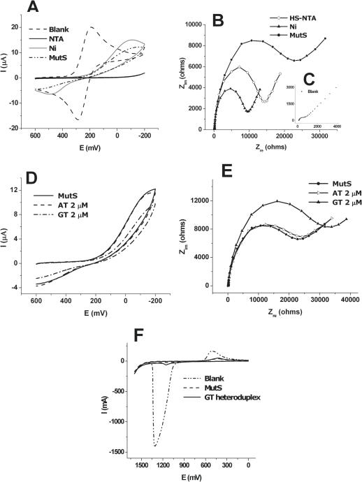
Figure 2.
(A) Cyclic voltammogram for blank, HS-NTA coated step, Ni-NTA coated step and MutS immobilized step in the presence of 5 mM . Indirect detection of the state of electrode was possible through observation of the change of redox peaks in CV. (B) Nyquist plots for each immobilized step, HS-NTA coated step (open diamond), Ni-NTA-HS coated step (closed triangle), MutS immobilized step (closed circle). After immobilization of HS-NTA, Rct is increased (open diamond), then formation of Ni-NTA complex causes a decrease of Rct. After immobilization of protein, Rct is re-increased due to interference degree of for the condition of electrode surface. (C) Blank in the presence of 5 mM . (D) Cyclic voltammogram and (E) Nyquist plots measured after the reaction of AT homoduplex and GT heteroduplex. The CV and impedance results shown in (D) and (E) are the specific interaction between MutS and heteroduplex (dash-dot line and closed triangle line), whereas indicated little change between MutS and homoduplex (dashed line and open triangle line). (F) Differential pulse voltammograms measured at 0 to +1.6 V before and after the reaction of GT heteroduplex. Differential pulse voltammetry (DPV) was used to observe the oxidation peak of adenines of the dsDNA captured by MutS after interaction of the MutS probe with GT heteroduplex.

