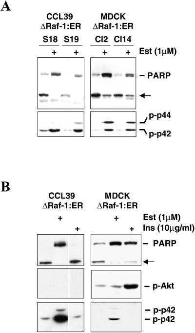
Figure 8.
Inhibition of PARP cleavage in CCL39- or MDCK-derived clones expressing ΔRaf-1:ER. (A) Western blot analyses of PARP cleavage (top) and MAP kinase activation (bottom) were performed on total cell lysates from two independent clones expressing ΔRaf-1:ER after 10 h of anchorage and serum withdrawal in the presence (+) or absence of 1 μM estradiol. Clones S18 and S19 (left) were derived from CCL39; clones Cl2 and Cl14 (right) were derived from MDCK. (B) Western blot analyses of PARP cleavage (top), Akt activation (middle), and MAP kinase activation (bottom) were performed on total cell lysates from CCL39–ΔRaf-1:ER cells (clone S18) and MDCK–ΔRaf-1:ER clone 14 (Cl14) after 10 h of anchorage and serum withdrawal in the presence (+) or absence of 1 μM estradiol or 10 μg/ml insulin (Ins).

